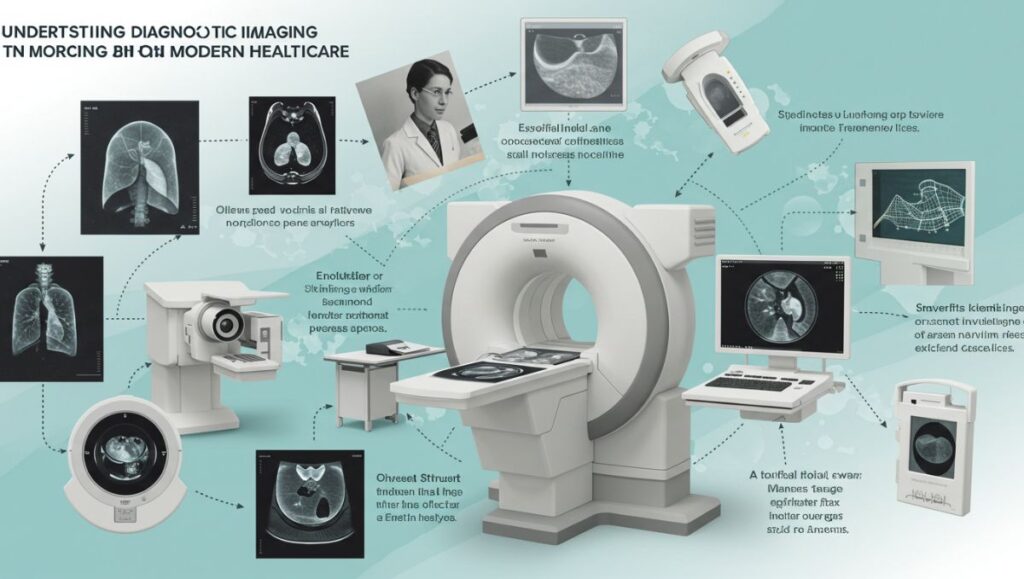
In modern medicine, few technologies have transformed healthcare as profoundly as diagnostic imaging. Often referred to as DIAG imaging, this field encompasses a wide range of techniques that allow doctors to see inside the human body without surgery. From simple X-rays to advanced MRI and CT scans, diagnostic imaging helps detect, monitor, and treat countless medical conditions.
Today, DIAG image technologies are indispensable tools in hospitals, clinics, and research centers worldwide. They allow physicians to make accurate, timely diagnoses, improve patient outcomes, and reduce the need for invasive procedures.
This article explores what DIAG imaging is, how it works, its various types, and why it remains one of the cornerstones of modern healthcare.
What Is a DIAG Image?
A DIAG image, short for diagnostic image, is a visual representation of the inside of the human body, produced using medical imaging technologies. These images reveal structures such as bones, organs, tissues, and blood vessels, enabling doctors to detect abnormalities, injuries, and diseases.
In essence, DIAG imaging provides non-invasive access to the body’s internal systems. Physicians use these images to:
-
Diagnose medical conditions accurately.
-
Monitor the progress of diseases.
-
Guide surgical or therapeutic procedures.
-
Evaluate treatment effectiveness.
Whether it’s identifying a broken bone or detecting cancer at an early stage, DIAG imaging plays a critical role in patient care.
How DIAG Imaging Works
The science behind diagnostic imaging involves using different forms of energy — such as X-rays, sound waves, radio waves, or magnetic fields — to produce detailed images of internal body structures. Each imaging method operates differently but shares the same goal: creating a visual map that helps medical professionals understand what’s happening inside the body.
Here’s a simplified explanation of how DIAG imaging works:
-
A device emits energy (like X-rays or sound waves) into the body.
-
These waves interact with internal structures (bones, tissues, fluids).
-
A detector or sensor captures the reflected or transmitted signals.
-
A computer converts these signals into digital images.
Doctors then analyze these images for abnormalities, injuries, or diseases.
Main Types of DIAG Image
DIAG imaging includes several major techniques, each suited for specific diagnostic purposes. Below are the most common types used in modern medicine:
1. X-Ray Imaging
X-rays are the oldest and most common form of diagnostic imaging. They use ionizing radiation to produce images of dense structures like bones and teeth.
Common Uses:
-
Detecting fractures, infections, or bone tumors.
-
Identifying lung diseases such as pneumonia.
-
Examining dental and orthopedic conditions.
Advantages:
-
Quick and inexpensive.
-
Widely available.
Limitations:
-
Limited detail for soft tissues.
-
Exposure to low levels of radiation.
2. Computed Tomography (CT Scan)
A CT scan, or CAT scan, uses rotating X-ray machines and computers to create cross-sectional images of the body. It provides more detail than a standard X-ray, allowing doctors to see internal organs, muscles, and blood vessels clearly.
Common Uses:
-
Diagnosing internal injuries and bleeding.
-
Detecting cancers and tumors.
-
Planning radiation therapy or surgeries.
Advantages:
-
Produces detailed 3D images.
-
Non-invasive and fast.
Limitations:
-
Involves higher radiation exposure than regular X-rays.
3. Magnetic Resonance Imaging (MRI)
MRI uses strong magnetic fields and radio waves to produce detailed images of organs and soft tissues. Unlike X-rays or CT scans, MRI doesn’t use radiation, making it safer for repeated use.
Common Uses:
-
Examining the brain, spinal cord, and nerves.
-
Evaluating joint and muscle injuries.
-
Detecting tumors and soft tissue abnormalities.
Advantages:
-
Highly detailed imaging of soft tissues.
-
No ionizing radiation.
Limitations:
-
Expensive and time-consuming.
-
Not suitable for patients with metal implants.
4. Ultrasound (Sonography)
Ultrasound imaging uses high-frequency sound waves to produce real-time images of organs and tissues. It’s commonly used in obstetrics to monitor pregnancies but has many other medical applications.
Common Uses:
-
Monitoring fetal development.
-
Examining the heart (echocardiography).
-
Assessing organs like the liver, kidneys, and gallbladder.
-
Guiding needle biopsies.
Advantages:
-
Safe and radiation-free.
-
Real-time imaging of moving organs.
Limitations:
-
Limited detail for structures behind bones or air-filled areas.
5. Positron Emission Tomography (PET Scan)
A PET scan uses radioactive tracers to visualize metabolic activity in the body. It shows how tissues and organs are functioning rather than just how they look.
Common Uses:
-
Detecting cancers and monitoring treatment response.
-
Evaluating brain function in Alzheimer’s disease.
-
Assessing heart muscle health.
Advantages:
-
Provides metabolic and functional information.
-
Often combined with CT or MRI for comprehensive imaging.
Limitations:
-
Exposure to small amounts of radioactive material.
-
High cost and limited availability.
6. Mammography
Mammography is a specialized form of X-ray imaging used to examine breast tissue. It’s one of the most effective tools for early detection of breast cancer.
Common Uses:
-
Routine breast cancer screening.
-
Diagnosing lumps or abnormalities in breast tissue.
Advantages:
-
Detects cancer early, improving survival rates.
-
Non-invasive and relatively quick.
Limitations:
-
May cause mild discomfort during compression.
-
Can occasionally yield false positives.
Applications of DIAG Imaging in Medicine
DIAG imaging is essential across nearly all branches of healthcare. Its applications range from simple fracture diagnosis to complex neurological studies.
1. Disease Detection
-
Identifies tumors, infections, and organ abnormalities.
-
Enables early detection of chronic conditions like cancer, heart disease, or diabetes complications.
2. Treatment Planning
-
Helps doctors plan surgeries, radiation therapy, or chemotherapy.
-
Provides real-time guidance during minimally invasive procedures.
3. Monitoring and Follow-Up
-
Tracks disease progression or regression during treatment.
-
Assesses the effectiveness of medications or therapies.
4. Emergency Medicine
-
Quickly identifies internal bleeding, fractures, or trauma after accidents.
5. Preventive Screening
-
Detects potential health problems before symptoms appear (e.g., mammograms, bone density scans).
Advantages of DIAG Imaging
-
Non-Invasive – Provides vital internal information without surgery.
-
Accurate and Fast – Delivers quick, reliable diagnostic results.
-
Early Detection – Helps find diseases in their earliest stages.
-
Guidance in Treatment – Improves precision during surgical and therapeutic procedures.
-
Improved Patient Outcomes – Leads to faster, safer recoveries.
Risks and Limitations of DIAG Image
While DIAG imaging offers enormous benefits, it also comes with a few considerations:
-
Radiation Exposure: Some techniques (like X-rays and CT scans) expose patients to small amounts of ionizing radiation.
-
Contrast Allergies: Some procedures use contrast dyes that may cause allergic reactions in sensitive individuals.
-
Cost and Accessibility: Advanced imaging (MRI, PET) can be expensive and limited in certain regions.
-
False Positives/Negatives: In rare cases, imaging results can be misleading, requiring additional tests.
Healthcare providers always weigh the benefits against potential risks before recommending any imaging procedure.
Technological Innovations in DIAG Imaging
The field of diagnostic imaging continues to evolve rapidly, driven by innovation and artificial intelligence (AI). Some of the latest advancements include:
1. AI-Assisted Image Analysis
AI algorithms can detect patterns in scans faster and sometimes more accurately than humans — improving diagnosis for conditions like cancer, stroke, or fractures.
2. 3D and 4D Imaging
New imaging systems provide 3D (and even 4D) views of organs in motion, allowing real-time analysis of how the body functions.
3. Hybrid Imaging Systems
Combining PET with CT or MRI creates comprehensive diagnostic tools that show both structure and function simultaneously.
4. Portable Imaging Devices
Compact ultrasound and X-ray machines are revolutionizing healthcare access in remote and emergency settings.
5. Cloud-Based Image Storage
Digital imaging systems now store DIAG images securely in the cloud, allowing doctors and patients to access them anytime, anywhere.
The Role of Radiologists in DIAG Imaging
Behind every DIAG image is a team of professionals ensuring accuracy and safety — particularly radiologists and radiologic technologists.
-
Radiologists are medical doctors who interpret diagnostic images, identify diseases, and collaborate with other physicians to plan treatments.
-
Radiologic Technologists (or imaging technicians) operate the imaging equipment, position patients, and ensure the quality of the scans.
Their expertise bridges technology and medicine, making DIAG imaging an essential part of patient diagnosis and care.
The Future of DIAG Imaging
The future of DIAG imaging lies in greater precision, personalization, and connectivity. Upcoming technologies like AI-driven image enhancement, quantum imaging, and wearable scanners promise to make diagnostics faster, safer, and more accurate.
Moreover, personalized imaging protocols based on genetic and molecular profiles will allow doctors to tailor imaging procedures to individual patients — ushering in a new era of precision medicine.
Conclusion
DIAG imaging represents one of medicine’s most remarkable achievements — a window into the human body that allows doctors to diagnose, treat, and prevent diseases with unprecedented accuracy.
From the simplicity of an X-ray to the sophistication of an MRI or PET scan, diagnostic imaging continues to redefine how medicine understands the body. As technology advances, DIAG imaging will only become more powerful, accessible, and essential in saving lives and improving healthcare worldwide.


